Full-size Scanning Electron Microscopes (SEMs) have been restricted in use to experienced Electron Microscopists for decades. The advent of Benchtop SEM in the last two decades made Electron Microscopy accessible to beginners. These small systems have gained popularity due to their affordable price, low running costs and ease of use.
Shrinking a full-size SEM that can occupy up to half a room to the size of a coffee machine that can sit on a tabletop was accompanied by compromises on performance, resolution, flexibility and accuracy. This is why Benchtop SEMs had been more suited for high school teaching or as sample screening tools prior to imaging with a full-size SEM in research institutes. However, the technology has advanced many folds in the recent years. Similar to a smartphone that delivers performance equivalent to a computer from some years ago, some of the Benchtop SEMs have come closer in performance to full size SEMs. Though the flexibility in terms of attachments and future expansion is still not at par with full size SEMs, the resolution and analytical accuracy are comparable.
With so many Desktop SEM options on the market and each one blaring their own sales pitch, making a decision can get confusing. More importantly the worry of making a wrong decision can become overwhelming. NanoTechnology Solutions offer this guide prepared from our research, academic, technological and maintenance experience with Electron Microscopes to assist Researchers in making calculated decisions.
Consider your needs
If the purpose of Benchtop SEM is teaching or to screen samples before analysing with a full-size SEM, any Benchtop SEM would do the job. Even a few years old system can quickly shortlist samples to reduce your paid beam-time on big SEM. It is advisable to pick the cheapest available model.
However, if there is no access to a full-size SEM or the aim is to become self-sufficient in most of the SEM work, not every Benchtop system will meet the standards due to the compromises mentioned earlier.
Resolution of Benchtop SEM
From experience, users always want more resolution out of their Benchtop SEM so it is a good practice to get the best resolution your budget allows.
Does the manufacturer state resolution on the brochure?
Some manufacturers do not mention the resolution of their Benchtop SEM on brochures. Full-size SEMs with tungsten filaments (i.e. non-Field Emissions type) offer a resolution of 3 nm. Today it is possible to purchase Benchtop systems with 5 nm resolution.
Accelerating Voltage on Benchtop SEM
Full-size SEMs offer Accelerating Voltage range from 0.3 kV to 30 kV in order to allow users to choose the right conditions for different samples (bio, polymers, metals, ceramics etc.)
Operating the SEM at low accelerating voltages (below 5 kV) is important in the observation of non-conductive charging samples, imaging of beam-sensitive samples and obtaining more surface information from conductive as well as non-conductive specimens.
Low energy of 1 or 2 kV could help investigate organic contamination on various substrates. Thin layers of organic contamination are electron transparent to higher voltages and the SEM would not be able to see them at high kV.
Some materials may be too sensitive to 15 kV energy but have low electron yield at 10 kV and other materials may even be sensitive to 10 kV and offer very low electron yield at 5 kV. In such cases, a voltage in between (e.g. 8 or 12 kV) is useful to balance between beam damage and signal to noise ratio.
Does the Desktop SEM offer this flexibility of precisely choosing the Accelerating Voltage or is restricted to 5, 10, 15 kV steps?
Why 30kV?
For decades, high Accelerating Voltages ranging from 15 kV to 30 kV have been used by Electron Microscopists for imaging and analysing materials because of the nanometer scale image resolution and the ability to excite the renowned K-lines in the EDS atomic spectra for the identification and accurate quantification of elements. This is critical for samples where materials with similar elemental compositions and/or containing elements with overlapping EDS energy peaks that can be misinterpreted due to misidentification of elements and/or inaccurate quantification.
For example, in the EDS spectrum below, L-line of Zr overlaps with K-line of P at ~2 kV. It would be difficult to know which element was causing a peak near 2 kV or whether both elements were contributing to the peak (and with what ratio). With the capability of running EDS analysis above 15kV, user would be able to see whether a peak shows up at ~15.75 kV, which is the K-line of Zr. Quantitative analysis would reveal percentage of P hidden in the Zr L-line peak based on intensity of the Zr K-line peak at ~15.75 kV.

Experts at ElementPi in the USA did a comparison of 15kV and 30kV EDS analysis. Their comments are mentioned below:
“The majority of tabletop SEMs are restricted to 15 kV maximum accelerating voltage for the electron beam. This limited accelerating voltage does not promote collecting satisfactory spectra in the range above 8-10 keV for many critical elements like Molybdenum, Bromine, Zinc and several others. Further, 15 kV forces the use of spectral peaks at energies less than 5 keV where there are frequently many overlapping elements. As shown in figure below, an electron beam with accelerating voltage up to 30 kV can generate better imaging and better EDS results by matching the beam to the composition of the sample.”

In-situ sample height adjustment
Scanning Electron Microscopes require a longer working distance for EDS analysis which compromises on resolution. In order to obtain high resolution SEM image of the same area, the SEM must have a capability to adjust sample height while the sample chamber is closed and the beam is ON. Most Benchtop SEMs require breaking the vacuum and taking the sample out to adjust the optimal Working Distance from SEM to EDS and vice versa. This results in the area of interest being lost and the user spending significant time to find the area of interest again.
Ideally the Benchtop SEM should have a capability to adjust the sample height in-situ.
Stage Control on Benchtop SEM
Most SEMs offer motorised stage control through the SEM software which is very convenient. Some offer joystick control in addition which does not really offer superior capability. Personal liking of the users would decide which control is their preferred method. Sometimes it can be beneficial to have both types of control to cater for diverse users. Motorised stage can help with high throughput and allow calling the XY coordinates of previously captured locations so that the areas previously imaged could be investigated again e.g. when the user needs to return to a location to conduct EDS analysis.
Spot Sizes
Different areas within a sample or different magnifications may demand change of beam intensity. For example, smaller spot sizes are good for high magnifications while larger spot sizes are better for low magnifications. For publishable quality images, it is important to have such flexibility.

The images can be dramatically improved with a simple click in the SEM software. However, beginners in SEM can use a fixed spot size setting and still use it as a basic Desktop SEM with no flexibility. A high-resolution Desktop SEM at fixed setting would still give better images than a basic system with fixed settings.
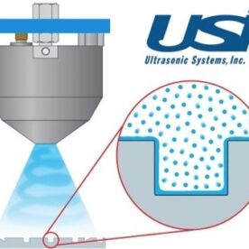
Moveable Aperture Strip on a Benchtop SEM?
Advanced SEMs come with moveable aperture strip with different sized apertures. Large size aperture provides greater signal for imaging and EDS work while small aperture provides very high resolution and depth of field. Users can move between different apertures by simply turning a knob and selecting the one delivering them the best result.
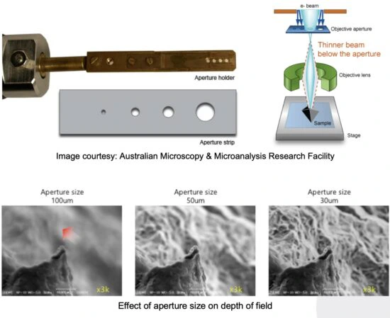
Images above show that by simply changing the aperture size, image quality can be significantly improved. This is highly beneficial for samples with large 3D features. Beginners in SEM can use one of the middle size apertures for all imaging and still get better images than a Benchtop SEM with single fixed aperture.
The aperture strip also helps in reducing downtime. When one aperture is dirty, users can just turn the knob and use the next one on the strip. Dirty apertures result in poor image quality and SEMs with a single fixed aperture have the potential of getting contaminated four times faster.
Imaging in Low Vacuum
Is the SEM supplier confusing the Low Vacuum or Environmental SE detector with an Environmental SEM (ESEM)? Environmental SEMs are specially designed and have complex vacuum systems that allow levels up to 4000 Pa while Benchtop SEMs use few 10’s of Pa at best.
For decades, it has been the industry standard that SEMs come with high vacuum SE imaging capability which is very important for high quality imaging. For low vacuum observation, uncoated samples and compositional information, BSE imaging is used.
Because of their geometry as shown below, SE detectors provide better topographical information (side view) as compared to a BSE detectors (top view).

This is the reason BSE images appear flatter than SE images. However, modern BSE detectors have four segments which allow enhanced topography by turning off one or two segments of the detector and give SE-like images.

The beauty of 4-segment BSE detectors is the capability of switching between COMPO (4 segments) and TOPO (2 or 3 segments) modes. SE detectors only offer topographical information. Some Benchtop SEMs offer low vacuum SE imaging which is good for uncoated samples that may charge under high vacuum. However, most BSE detectors provide this capability. Importantly, the absence of high vacuum SE imaging means the lack of high quality images.
Most SEMs live up to the industry standard and offer a high vacuum SE detector.
Cooling Stage in Desktop SEM
Cooling the sample can reduce thermal damage on sensitive samples. More importantly minimise moisture evaporation from samples and prolong viewing times.

Working Distance Range
Previously we learnt to improve the depth of field by selecting a smaller aperture. The depth of field can further be improved by increasing the working distance. Images below demonstrate the effect of combination of different aperture sizes and working distances.
NOTE: The working distance is not to be confused with the sample height.

Focal length of the objective lens of the SEM would determine the ultimate depth of field it could deliver.
Is the Benchtop SEM able to focus samples at 30 mm or 40 mm working distance?
Pixels of Benchtop SEM images
Images can show more detail on a projector and can be printed as posters if they have higher pixel density. Does the SEM allow image resolution of up to 5120 x 3840 or is limited to 2560 x 1940 or 1280 x 960?
Low pixel images could show jagged edge artefact known as pixilation when projected or printed in journals. Higher pixel density can be useful for producing a high-quality image with rich topography and clean, well-defined edges.
Magnification of Benchtop SEM
Magnification can be misleading as it is a limit set by the software. What is more important is the resolution of the electron optics. For example, ‘Tabletop SEM A’ with 30 nm resolution and 300,000x magnification would perform much worse than ‘Tabletop SEM B’ with 3 nm resolution and 100,000x magnification. Tabletop SEM A would show blurry images even at 50,000x as compared to Tabletop SEM B because of the difference in capability of electron optics i.e. the hardware.

Users should be careful of ‘digital zoom’ or ‘monitor magnification’ specifications as they are not true representation of the magnification power of the electron optics.
Beginner and expert software versions
All Desktop SEMs offer simple and easy to understand software which is immensely useful to bring beginners up to speed.
If the SEM offers an expert version that allows controlling fine settings that the advanced user can change if required, it could give greater flexibility and cater for the needs of occasional and expert users alike.
Pre-Centred Tungsten Filament
Most Benchtop SEMs today offer easy maintenance and easy filament replacements by using pre-centred filament cartridges.
Spare part lead time
How quickly can the supplier send spare parts during breakdown? While filaments are generally kept in stock, some Electron Microscope manufacturers take over 3 months to ship out spare parts.
Applications Support
Does the supplier have a true Electron Microscopist with years of hands-on experience handling a diversity of samples? Often times companies give the designation of ‘Applications Specialist’ to someone who occasionally used the SEM for a university project and use it as a misleading sales tactic.
Someone with demonstrated experience can help you make the most out of your SEM by resolving issues with the samples or operating conditions and help you publish great images.
Tender requirements
If the purchase is going to tender, it is a good practice to write generic specifications that allow everyone to bid. This demonstrates the buyer is fair and helps get competitive pricing from all bidders. Buyers should be wary of sales people suggesting to put ‘lock-out specifications’ in the tender as these are meant to lock you into buying their system at higher price than a competitive situation. Other bidders can be put off by this and may refuse to bid or complain of unfair procurement process.
Generic specifications allow everyone to bid at their best offers. Suppliers lose tenders all the time and understand only one can win in the end however if they see no chance of winning even before they bid, this can hurt the competitive process.
While evaluating different bids, try to stay focused on things that affect your end goal i.e. the SEM results and avoid distractions that do not directly improve the end goal.